Microscopy has revolutionized the field of biology, allowing scientists to explore the intricate world of cells and tissues with unprecedented detail. However, traditional microscopy techniques have limitations when it comes to imaging thick biological samples, which can distort light and create blurry images. In an effort to address this issue, researchers at HHMI’s Janelia Research Campus have turned to techniques used in astronomy to enhance the clarity and sharpness of microscopy images.
Astronomers have long used methods to correct for distortions caused by the atmosphere, allowing them to capture clearer images of far-away galaxies. By applying similar techniques to microscopy, such as adaptive optics, researchers can improve the quality of images of biological samples. However, traditional adaptive optics methods are complex, expensive, and slow, making them inaccessible to many labs. To make these techniques more widely available to biologists, the team at Janelia Research Campus has explored the use of phase diversity methods, which have been successful in astronomy but are relatively new to the life sciences.
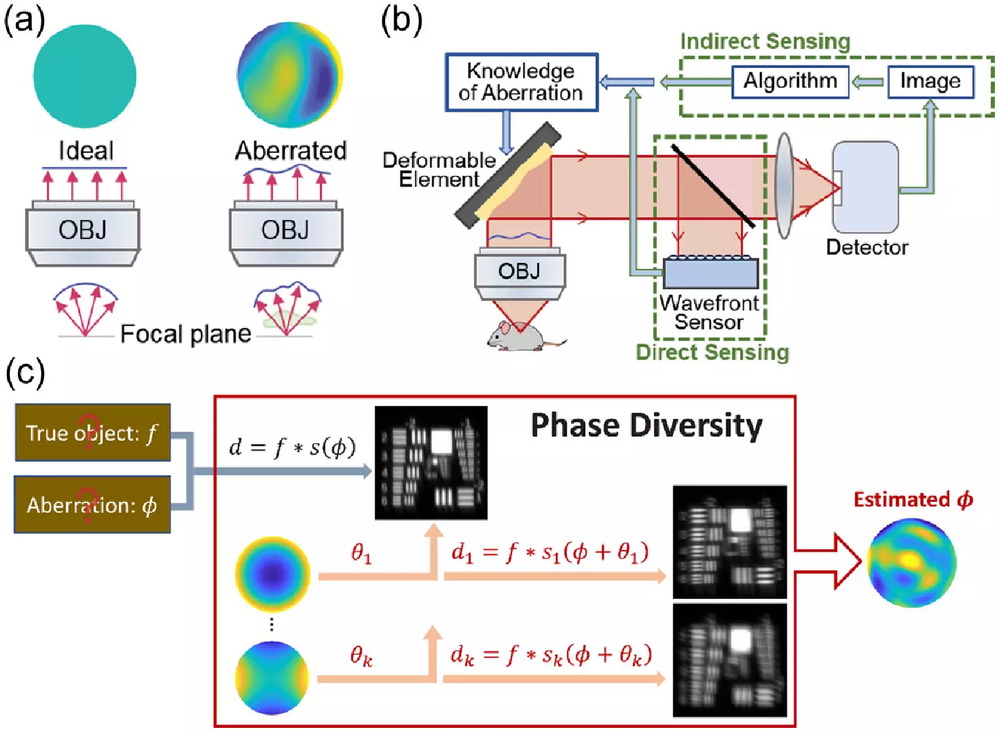
Phase diversity methods involve adding images with known aberrations to a blurry image with unknown aberrations, allowing for the unblurring of the original image. Unlike other adaptive optics techniques, phase diversity does not require significant modifications to existing imaging systems, making it a cost-effective and efficient solution for microscopy. The team first adapted the astronomy algorithm for microscopy and tested it with simulations. They then enhanced a microscope with a deformable mirror and additional lenses to create known aberrations, improving the software used for phase diversity correction.
In testing their new method, the team found that they could calibrate the microscope’s deformable mirror significantly faster compared to existing methods. They also demonstrated the ability of the new technique to sense and correct randomly generated aberrations, resulting in clearer images of fluorescent beads and fixed cells. The next phase of their research will involve testing the method on real-world samples, such as living cells and tissues, and expanding its application to more advanced microscopy systems. The team also aims to streamline the method to make it more automated and user-friendly.
The development of this new method represents a significant advancement in microscopy, offering a faster and more affordable way to implement adaptive optics techniques in biological imaging. By making these techniques more accessible to a wider range of labs, researchers hope to enhance the clarity and detail of microscopic images, enabling biologists to gain deeper insights into the intricate world of cells and tissues. As the technology continues to evolve, the future of microscopy holds the promise of even greater discoveries and breakthroughs in the field of biology.
Leave a Reply