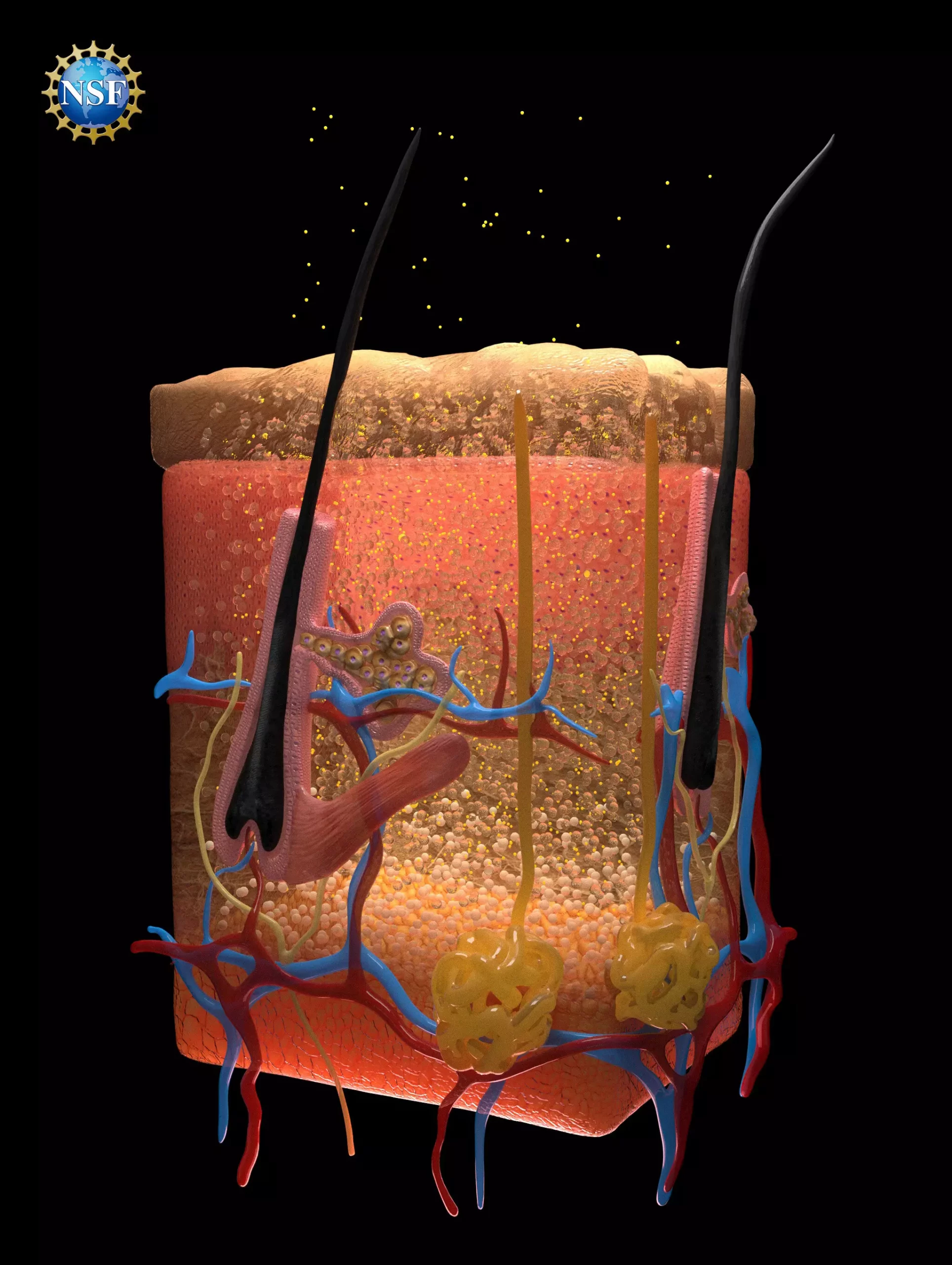
A groundbreaking technique recently developed by researchers at Stanford University promises to transform the way we visualize internal organs, potentially enhancing a myriad of medical diagnostics. The study, titled “Achieving optical transparency in live animals with absorbing molecules,” was published in the September 6, 2024, issue of Science. The research showcases the ability to render biological tissues transparent using a food-safe dye, which may have profound implications for diagnosing injuries, monitoring digestive disorders, and identifying cancers.
What sets this innovation apart is its counterintuitive approach: it involves the application of a dye that is not only accessible but also reversible, making it suitable for various medical applications. Assistant Professor Guosong Hong, one of the lead authors, envisions that this technique can improve clinical practices, such as blood drawing and laser treatments for tattoo removal and cancer therapy.
The core of this technique lies in the researchers’ in-depth understanding of light interactions with biological tissues. They recognized that the scattering of light within our bodies is due to different materials—like fats, proteins, and fluids—each possessing unique refractive indices. When light passes through these varying materials, it scatters, rendering our tissues opaque.
To counteract this issue, the research team aimed to equalize the refractive indices of biological materials. They discovered that specific dyes, particularly tartrazine (FD & C Yellow 5), effectively absorb light while facilitating a uniform passage of light through tissues. The decisive factor was the dye’s ability to align the refractive indices of the fluids within muscle cells, enabling these cells to become transparent when the dyestuff was adequately absorbed.
The researchers began their journey by investigating how microwave radiation interacts with biological tissues, which led them to rediscover certain optical principles outlined in the literature dating back to the 1970s and 1980s. Key concepts such as Kramers-Kronig relations and Lorentz oscillation provided foundational knowledge to understand how specific dyes could alter refractive indices of biological fluids.
This exploration transitioned smoothly from theoretical groundwork to practical experimentation. Initial tests involved applying the tartrazine solution to animal subjects, revealing a dynamic view of their circulatory system and organ movements in real time. Notably, the solution, once rinsed off, allowed tissues to swiftly return to their normal opacity without adverse effects, indicating that the application was both temporary and safe.
The potential applications of this technology extend far beyond mere visualization. For example, enhancements in laser-based procedures could lead to more effective treatment modalities for cancerous cells situated deeper in the body. Moreover, the prospect of using this technique to monitor digestive health and identify anomalies in various organ systems opens a new chapter in utilizing optical technologies for health advancements.
The researchers hypothesize that injecting the dye may enable even deeper views within organisms, bridging the gap between microscopic and macroscopic imaging. This could lead to quicker and more accurate diagnoses, significantly improving patient care and reducing the need for invasive procedures.
The success of this program relied heavily on collaboration among a team of 21 members that included students, researchers, and advisors, each contributing their expertise. An unexpected hero in this evolution was an old yet functional ellipsometer found in the Stanford Nano Shared Facilities. Traditionally used in semiconductor manufacturing, the ellipsometer demonstrated that even fundamental tools can yield groundbreaking insights when applied creatively in a different context.
Richard Nash, an NSF Program Officer, emphasized the importance of access to varied instrumentation, stating that sometimes older technology can catalyze innovative discoveries in unexpected fields, as illustrated by this project.
The advancement presented by these Stanford researchers represents a pivotal moment in the intersection of optics and medicine. By leveraging fundamental principles from physics, they have not only provided a novel method for achieving organ visibility but have also laid the groundwork for a new area of study aimed at matching dyes to biological tissues based on optical properties.
As noted by Adam Wax, another NSF Program Officer, the implications of exploiting well-understood equations to develop transformative applications in the medical field are profound and exciting. As research continues, this technology might very well become a standard practice in medical diagnostics, promising a future where understanding human physiology is as vivid as seeing through clear glass.


Leave a Reply