The exploration of neural circuits has become increasingly sophisticated with the advent of genetically encoded voltage indicators (GEVIs). These innovative tools enable researchers to visualize the electrical activity of neurons, a pivotal aspect of understanding how neuronal communication and information processing occur in the brain. Despite their promising applications, the debate surrounding the efficacy of one-photon (1P) versus two-photon (2P) voltage imaging continues to generate considerable interest and discussion within the scientific community.
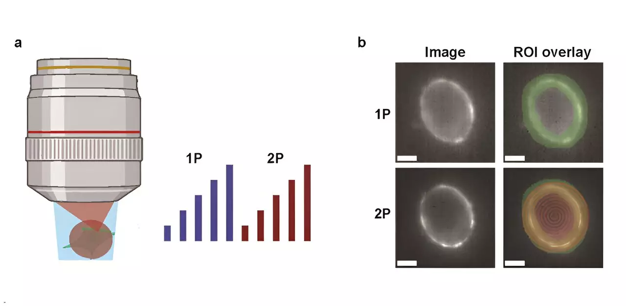
Recent research conducted at Harvard University provides crucial insights into the advantages and drawbacks inherent to 1P and 2P imaging techniques. The study, published in the journal Neurophotonics, meticulously contrasts the optical properties and biophysical limitations of the two methods. By evaluating the brightness and voltage sensitivity of several commonly used GEVIs under 1P and 2P illumination, the researchers were able to establish a clearer understanding of how each technique functions in practical applications.
A major aspect of their investigation revolved around how fluorescence intensity diminishes with depth in mouse brain tissue. As in vivo imaging increasingly requires exploration of deeper brain regions, this parameter is critical in determining the feasibility of both methods. The study benefitted from the development of a predictive model capable of determining the number of neurons that can be feasibly measured, factoring in the characteristics of voltage reporters, imaging configurations, and desired signal-to-noise ratios (SNR).
One of the standout findings from the study is the staggering illumination power requirement for 2P excitation, which is approximately 10,000 times greater per neuron compared to 1P. This immense power demand introduces multifaceted challenges, such as risk of tissue photodamage and increased shot noise, potentially obscuring the data collected during imaging. For example, in trials involving the JEDI-2P indicator positioned within the mouse cortex while targeting an SNR of 10, the research indicated that it would be feasible to capture data from only about 12 neurons at depths that exceed 300 micrometers, even with optimized laser settings.
The results of the Harvard study underscore the inherent trade-offs between 1P and 2P voltage imaging in terms of depth penetration and obtainable signal clarity. The limitations on current GEVIs suggest that further advancements in two-photon voltage imaging for in vivo applications will hinge on either significant enhancements in the technology of the sensors themselves or perhaps the exploration of entirely new imaging strategies.
The insights garnered from this critical evaluation not only illuminate the complexity of imaging neural activity but also highlight the imperative for continuing innovation within this vital field of research. As scientists seek to understand the intricacies of neural circuits, this study serves as a foundational reference that will inform future technological developments and methodologies.
Leave a Reply