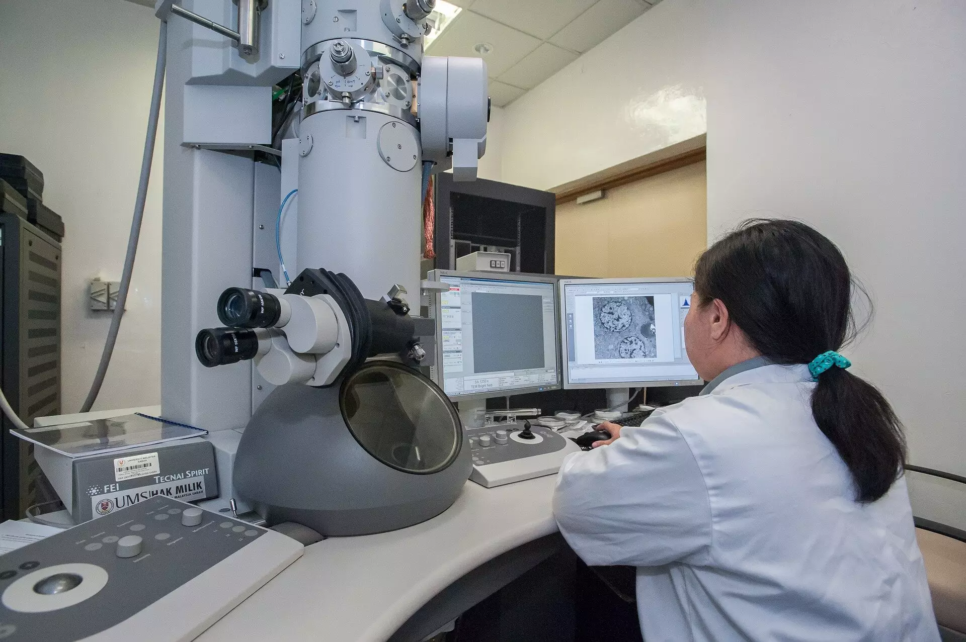
Cutting-edge advancements in scientific imaging have the potential to transform numerous fields, from molecular biology to materials science. An international team of researchers, led by Trinity College Dublin, has recently developed an innovative microscopy technique that significantly minimizes the time and radiation exposure necessary for effective imaging. By leveraging state-of-the-art technologies, this new method stands to enhance the quality of images captured while preserving the integrity of sensitive samples that are often vulnerable to radiation-induced damage. The implications of this breakthrough are vast, touching upon crucial areas such as biological research, where the pristine condition of samples is imperative for accurate analysis.
The Limitations of Traditional Methods
Traditionally, scanning transmission electron microscopes (STEMs) have relied on a uniform exposure strategy, where a focused beam of electrons is meticulously directed across a sample. The conventional approach dictates a fixed dwell time for each pixel, leading to a uniform level of radiation exposure across various regions of the image. While this simplistic method is easy to implement, it poses significant risks; excessive radiation can cause irreversible damage to delicate biological specimens. This uniform exposure method can yield results that are not only time-consuming but also detrimental, as research ultimately hinges on the quality and reliability of the images obtained.
Reimagining Imaging Logic
The pioneering work by the Trinity College team revolved around rethinking how imaging processes operate. Rather than adhering to fixed timing protocols, they introduced an event-based detection system, capable of gauging the time taken to detect a specific number of events, or electron interactions. This radical shift in approach permits microscopists to achieve the same image quality while using a fraction of the traditional radiation levels. The novel mathematical framework posits that the initial electron captured at each probing site provides significant information, whereas additional electron hits yield diminishing returns concerning image quality. This insight allows researchers to optimize the imaging process, radically reducing the risk of damage to specimens while also improving efficiency.
The Tempo STEM Patented Technology
Regardless of the sound theoretical basis behind their innovation, real-world application hinges on the development of tangible technology. The team unveiled the Tempo STEM, a patented system that integrates a high-tech “beam blanker.” This mechanism allows researchers to precisely control the electron beam, turning it off once optimal imaging conditions have been met. According to Dr. Lewys Jones, a key contributor to this research, the ability to modulate the electron beam in real time marks an unprecedented advancement in microscopy capabilities. With the Tempo STEM, researchers can achieve high-resolution images while dramatically lowering the radiation dose, thus enhancing the safety and integrity of sensitive biological samples.
Impact on Biological Research
Perhaps one of the most exciting aspects of this breakthrough is its direct application to biological microscopy. As noted by Dr. Jon Peters, biological samples are often significantly altered or destroyed under heavy radiation exposure, leading to either unusable images or misinterpretations of the data. Given that electrons in a microscope travel at astonishing speeds, the potential for damage remains a pressing concern. The introduction of the Tempo STEM not only alleviates these concerns but also enhances capability, offering a more reliable means of acquiring data.
Future Prospects and Innovations
As this innovative imaging method sets a new precedent, the excitement within the scientific community is palpable. Beyond biology, applications in materials science and technology are equally promising. Improved imaging could facilitate the exploration of new materials, aiding innovations in industries ranging from electronics to renewable energy. The versatility of this technique opens doors previously thought closed, empowering researchers to explore both known and uncharted territories within their respective fields.
This moment represents not just a technological advancement but a paradigm shift in how we perceive and conduct scientific imaging, positioning researchers to ask more ambitious questions and pursue deeper insights into the natural world. As the scientific community embraces this groundbreaking method, the landscape of microscopy—and the knowledge it yields—stands on the brink of a vibrant new era.


Leave a Reply