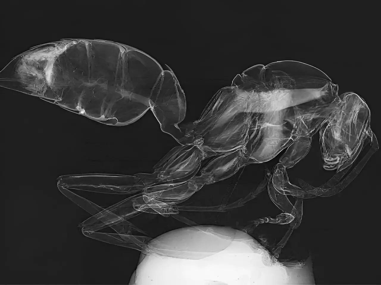
The groundbreaking advancements in X-ray imaging technology unveiled by researchers at the University of Houston have the potential to revolutionize various fields such as medical diagnostics, materials, industrial imaging, and transportation security among others. The introduction of a novel light transport model for a single-mask phase imaging system by Mini Das, Moores professor at UH’s College of Natural Sciences and Mathematics, along with Jingcheng Yuan, a physics graduate student at UH, promises significant improvements in non-destructive deep imaging for a wide range of applications.
Traditional X-ray technology heavily relies on X-ray absorption to generate images, which often leads to challenges when dealing with materials of similar density. This results in low contrast images and difficulties in distinguishing between different types of materials. These limitations have posed significant challenges across various fields including medical imaging, explosive detection, and beyond.
In recent years, X-ray phase contrast imaging (PCI) has gained considerable attention due to its ability to provide enhanced contrast for soft tissues by leveraging relative phase changes as X-rays pass through an object. Among the various techniques available, the single-mask differential technique stands out for its simplicity and effectiveness in practical applications, offering higher contrast images compared to other methods. The use of a single-shot, low-dose imaging method further streamlines the process.
The innovative light transport model developed by Das and his team aims to enhance the understanding of contrast formation and the interaction of multiple contrast features within acquired data. This enables the retrieval of images with two distinct types of contrast mechanisms from a single exposure, marking a significant advancement over conventional methods. By using an X-ray mask with periodic slits, the design creates a compact setup that boosts edge contrast and simplifies the overall process.
The research team has rigorously tested the model through simulations and on their in-house developed X-ray imaging system. The next phase involves integrating this technology into portable systems and adapting existing imaging setups to assess its performance in real-world scenarios such as hospitals, industrial X-ray imaging, and airport security. The simplicity, effectiveness, and cost-efficiency of this approach make it a versatile solution for enhancing image contrast and addressing critical needs in non-destructive deep imaging.
The innovative approach to X-ray imaging technology introduced by the University of Houston researchers has the potential to reshape the landscape of medical diagnostics, materials imaging, and security screening. By simplifying the setup, improving image contrast, and enabling single-shot imaging, this new technology opens up new possibilities in the field of X-ray imaging, making it more accessible, practical, and effective for a wide range of applications.


Leave a Reply