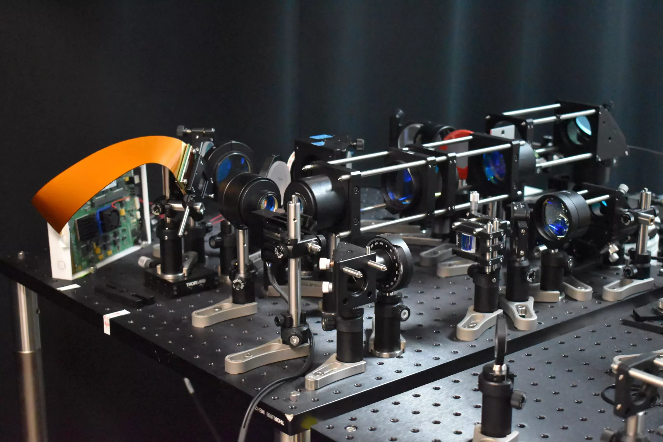
Neuroscience research has taken a significant leap forward with the development of a new two-photon fluorescence microscope. This cutting-edge technology allows for the rapid capture of high-speed images of neural activity at cellular resolution. Unlike traditional two-photon microscopy, this innovative approach minimizes harm to brain tissue while providing a clearer understanding of how neurons communicate in real-time. Lead researcher Weijian Yang from the University of California, Davis, emphasizes the importance of studying the dynamics of neural networks to gain insights into fundamental brain functions such as learning, memory, and decision-making. By using this new microscope, researchers can observe neural activity during various processes, shedding light on the intricate communication among neurons.
The Technology Behind the Microscope
The newly developed two-photon fluorescence microscope introduces a novel adaptive sampling scheme and replaces point illumination with line illumination. This method allows for in vivo imaging of neuronal activity in a mouse cortex at speeds ten times faster than conventional two-photon microscopy, with a reduction in laser power on the brain by more than tenfold. The adaptive sampling scheme targets active neurons using a digital micromirror device (DMD), which shapes and steers a short line of light to illuminate specific areas of the brain. Furthermore, a technique was devised to mimic high-resolution point scanning, enabling the reconstruction of detailed images for identifying regions of interest quickly. These advancements combine to create a powerful imaging tool that facilitates the study of dynamic neural processes in real-time while reducing damage to living tissue.
Implications for Neuroscience Research
The application of this new microscope extends to studying the pathology of neurological diseases at their earliest stages. By capturing calcium signals, indicators of neural activity, at a speed of 198 Hz in living mouse brain tissue, researchers can monitor rapid neuronal events that might be missed by slower imaging methods. Moreover, the ability to isolate the activity of individual neurons enables a more precise interpretation of complex neural interactions and a deeper understanding of the brain’s functional architecture. Future developments aim to integrate voltage imaging capabilities into the microscope to provide a direct and rapid readout of neural activity. Additionally, the research team plans to utilize this technology for real neuroscience applications, such as observing neural activity during learning and investigating brain activity in disease states. Enhancements to the microscope’s user-friendliness and size reduction are also on the agenda to improve its utility in neuroscience research.
The development of the two-photon fluorescence microscope marks a significant advancement in neuroscience research. By providing real-time imaging of neural activity with minimal harm to living tissue, this technology opens up new possibilities for studying brain function and understanding neurological diseases. With further innovations and applications on the horizon, researchers are poised to make groundbreaking discoveries in the field of neuroscience using this cutting-edge tool.


Leave a Reply